The science of “brain mapping”
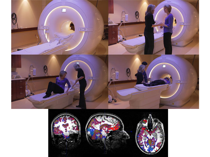
Brain scanning isn’t simple. It’s not like taking a photograph of your beloved in the park and instantly seeing their glowing face on your iPhone. There are more cells in the brain than stars in the Milky Way. Over 100 billion of them. And the scanner can collect data on all of them—using what is known as functional Magnetic Resonance Imaging, or fMRI. The scanner can catch people thinking, and localize some emotion and drive functions.
fMRI is a remarkable feat of modern technology. Who would have thought, even 25 years ago, that we would be able look into the minds of healthy, awake human beings and record what their brains are doing as they think. Moreover, it’s safe. Nothing touches the brain or body; nothing hurts; nothing damages our precious thinking or bodily processes.
And it operates on an intriguing principle: the harder brain cells are working, the more oxygen and glucose they demand to do their job, thus increasing blood flow to that area. The fMRI scanner is a huge magnet that records micro-changes in blood flow by tracking the minute traces of iron in your red blood cells.
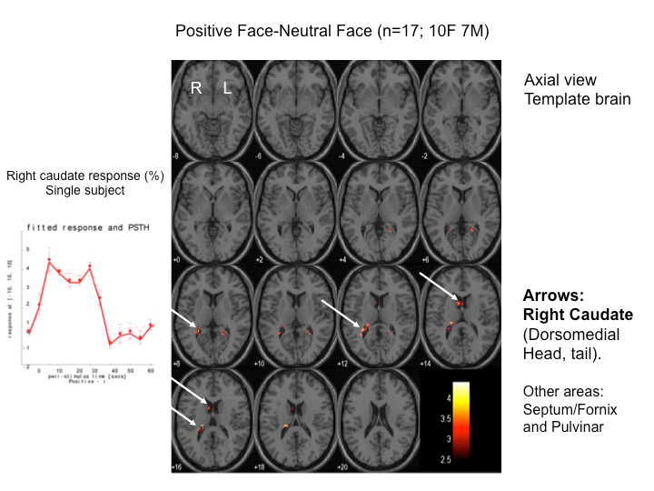
We use the words “activation” and “deactivation” to describe what happens. There are increases (activation) or decreases (deactivation) in blood flow to millimeter-sized areas of the brain. The amount of activation or deactivation depends on energy demands from all types of brain cells in each region. However, activation and deactivation are always relative to a control condition, and to a baseline that is an average of overall brain activity.

Enlightening!