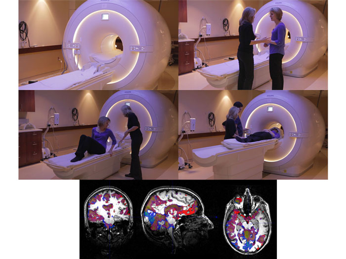
The scanning produces two types of images: 1) an anatomical image that shows the brain structure, 2) a functional image that shows the blood flow.
Placing the functional images (collected as the participant is “functioning” or doing something) on top of the anatomical images, Lucy can pinpoint those areas of the brain that are working.
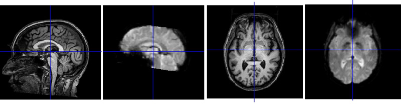
The far left image is an anatomical scan. Note that the brain is facing left, with the nose pointing left.
The second image is a “functional scan”—showing the brain regions that are demanding oxygen and glucose as they work. The blue crosshairs show the scientist where the computer estimates the anatomical and functional scans line up.
The two images to the right show anatomy and function again, but the slice through the brain is horizontal rather than from the side.
NEXT